The Evolution of Microscopy: From Hooke to Electron Microscopes
Share
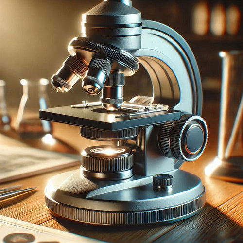
What if the biggest discoveries in science didn’t come from colossal telescopes peering into the cosmos but from tiny glass lenses looking inward? Imagine uncovering a hidden universe, teeming with life, right under your nose—literally. That’s the magic of the microscope.
Before microscopes, the microscopic world was a complete mystery. However, in the 17th century, a group of curious minds cracked open the door to the microscopic world, giving humanity its first glimpse of life too small to see with the naked eye. Today, microscopes allow us to dive deeper than ever before—right down to the building blocks of matter.
The Beginning: Hooke and the Birth of Cells
In 1665, Robert Hooke, an English scientist and inventor, published a book that would change science forever. In Micrographia, Hooke shared detailed sketches of what he saw under his handmade microscope, including the intricate structure of a cork. He noticed tiny, hollow compartments, like honeycombs, and named them “cells.” He didn’t know it then, but Hooke had just laid the foundation for cell biology—the study of life at its most basic level.
Hooke wasn’t alone, though. Across Europe, curious tinkerers and scientists were building their microscopes. One of them, a Dutch draper named Antonie van Leeuwenhoek, took things to another level.
Leeuwenhoek: The First Microbial Explorer
Antonie van Leeuwenhoek wasn’t a scientist by trade. He sold fabric. However, his obsession with grinding lenses and building microscopes led him to a groundbreaking discovery in 1674. Using a microscope smaller than a smartphone, van Leeuwenhoek peered into a droplet of pond water and saw tiny creatures he called “animalcules.” These were the first bacteria ever seen by human eyes.
Van Leeuwenhoek’s observations sparked a revolution. Suddenly, the world of the unseen was visible: microbes, blood cells, and sperm cells. His letters to the Royal Society of London, filled with detailed sketches, ignited interest and laid the groundwork for microbiology.
The Compound Microscope: Seeing Better and Bigger
Fast forward to the 19th century, when microscopes got an upgrade. Compound microscopes, which use multiple lenses to magnify objects, became the standard. This innovation allowed scientists to explore living tissues. This led to the development of cell theory, the idea that all living things are made of cells.
Around the same time, microscopes were used to study diseases. Robert Koch, a German physician, used a microscope to identify the bacteria that cause anthrax and tuberculosis, forever linking microbiology to medicine.
The 20th Century Breakthrough: Electron Microscopy
As powerful as compound microscopes were, they had limits. Light waves can only magnify so much, and scientists wanted to go deeper. Enter the electron microscope.
In 1931, two German scientists, Ernst Ruska and Max Knoll, developed the first electron microscope. Instead of light, this microscope used beams of electrons to illuminate specimens. This resulted in magnification power one thousand times greater than traditional microscopes. Scientists could now see viruses, organelles inside cells, and even individual atoms.
There are two types of electron microscopes: transmission electron microscopes (TEM), which provide detailed images of thin samples; and scanning electron microscopes (SEM), which create 3D images of surfaces. These tools opened up entirely new fields of study, from virology to nanotechnology.
Modern Microscopes: From Lasers to AI
Today’s microscopes are a far cry from Hooke’s handmade lenses. Advances in technology have made it possible to study living cells in real time, thanks to fluorescence microscopy. Scientists use fluorescent dyes to highlight specific structures inside cells, making them glow under ultraviolet light.
Even more futuristic are super-resolution microscopes, which can bypass the limits of light to see details as small as twenty nanometers. And let’s not forget artificial intelligence (AI). AI-powered software now helps scientists analyze complex microscope images faster than ever, accelerating discoveries in microbiology, medicine, and beyond.
Why It Matters:
Microscopes have shown us what life looked like billions of years ago in fossilized cells, and they’re helping us predict the future by studying the behavior of cancer cells, viruses, and bacteria. If the idea of peering into hidden worlds excites you, there are plenty of career options to explore:
- Biomedical Researcher: Study cells and microbes to find cures for diseases.
- Nanoengineer: Design and build materials at the molecular level.
- Forensic Scientist: Use microscopes to analyze crime scene evidence.
Fields like virology, pharmacology, and even art restoration rely heavily on microscopes, making it a versatile tool for almost any scientific passion.
From Hooke’s first glimpse of a cork to the super-resolution images of today, microscopes have changed how we see the world. They’ve revealed that life is more complex, beautiful, and interconnected than we could ever have imagined.
Additional Information:
Science Learning Hub. 2016. “History of Microscopy – Timeline.” Science Learning Hub. September 30, 2016. https://www.sciencelearn.org.nz/resources/1692-history-of-microscopy-timeline.
Ferlier, Louisiane. 2020. “Micrographia Online | Royal Society.” n.d. Royalsociety.org. https://royalsociety.org/blog/2020/07/micrographia-online/.
Kutschera, Ulrich. 2023. “Antonie van Leeuwenhoek (1632–1723): Master of Fleas and Father of Microbiology.” Microorganisms 11 (8): 1994. https://doi.org/10.3390/microorganisms11081994.
Lakhtakia, Ritu. 2014. “The Legacy of Robert Koch : Surmise, Search, Substantiate.” Sultan Qaboos University Medical Journal 14 (1): 37–41. https://doi.org/10.12816/0003334.